Research Article | | Peer-Reviewed

In Vitro Endothelial Differenthelial Assessment on Polyglycerol Sebacate / Polycaprolactone /Gelatin Electospun Scaffold

Received: 27 December 2024     Accepted: 11 February 2025     Published: 29 May 2025
Views:       Downloads:
Abstract

Due to the prevalence of cardiovascular diseases and their growing trend throughout the world, including Iran, efforts to treat these types of diseases have increased. There are lots of therapies for restoring cardiovascular function include organ transplants, reconstructive surgery, the use of mechanical or artificial devices, and the use of metabolic products. Although these methods are commonly used, they cause problems due to donor limitations, such as biocompatibility, infection, and tissue rejection by the patient. Meanwhile, vascular tissue engineering with the aim of building biocompatible and efficient vessels to replace lost vessels has created high hopes for the treatment of lesions. In this article, we used polyglycerol sebacate polymer (PGS) due to the properties such as high biocompatibility, good cell adhesion, controllable degradation rate and desirable mechanical properties, and combined it with polycaprolactone polymer (PCL) and gelatin to fabricate 3D scaffolds by electrospinning method. We also added vascular endothelial growth factor and then analysed the endothelial differentiation of mesenchymal stem cells. The expression of CD31 and VEGF-R2 genes has been measured by qPCR method, which revealed reasonable results.

Published in American Journal of Chemical and Biochemical Engineering (Volume 9, Issue 1)
DOI 10.11648/j.ajcbe.20250901.13
Page(s) 38-47
Creative Commons

This is an Open Access article, distributed under the terms of the Creative Commons Attribution 4.0 International License (http://creativecommons.org/licenses/by/4.0/), which permits unrestricted use, distribution and reproduction in any medium or format, provided the original work is properly cited.

Copyright

Copyright © The Author(s), 2025. Published by Science Publishing Group

Keywords

PGS, PCL, Gelatin, Stem Cells, Differentiation, Endothelial Differentiation, Electrospinning, In Vitro

1. Introduction
Given the prevalence of cardiovascular diseases and the increasing trend of myocardial infarction in developing countries, which causes permanent and irreversible cell death and destruction in a part of the heart muscle (myocardium), we realize the need to take steps towards treating these deficiencies. Given the increasing demand for organ transplantation and the shortage of organs and tissues (Organ Procurement and Transplantation Network, 2014), tissue engineers have turned to the development of synthetic analogues. This approach involves the proliferation of cells from a small number of stem cells and then culturing them in temporary 3D scaffolds to form new tissue or organs. In the meantime, vascular tissue engineering, with the aim of constructing biocompatible and efficient vessels to replace lost vessels (such as coronary arteries during a heart attack), has created great hopes for the treatment of these lesions. Traditionally, vascular tissue engineering has used co-culture of vascular smooth muscle cells (SMCs) and endothelial cells on collagen scaffolds, degradable scaffolds, or decellularized vessels . Since access to endothelial cells and smooth muscle cells (SMCs) requires sacrificing existing blood vessels, stem cells have gained a special place in vascular tissue engineering due to their easier accessibility and ability to proliferate and differentiate into various cell types, including endothelial cells and smooth muscle cells (SMCs), both inside and outside the body . A blood vessel has a highly porous fibrous layered structure with cells with different phenotypes in each layer. Among these different cells, endothelial cells have been the focus of many studies
For endothelial culture and differentiation, both embryonic stem (ES) cells and adult stem cells, which are extracted from different sources (such as mesenchymal stem cells, hematopoietic stem cells, and skeletal satellite myoblasts), have been used . Among them, mesenchymal stem cells are the most promising option in the field of cell therapy due to their ease of isolation and culture, as well as their ability to infiltrate and their immune properties. Although MSCs from different sources have common features, they also show important differences. These differences may indicate the unique local properties of the cavities from which the cells originate . Accordingly, in this study, adult bone marrow-derived mesenchymal stem cells were used. In addition to the type of stem cell selected, the biochemical and biomechanical environment also has a significant effect on stem cell differentiation . From this perspective, the type of culture medium and the types of physical and chemical stimuli can be considered the most important factors. The superiority of cell culture on 3D polymer scaffolds compared to other methods (including cell culture on plates) has been proven in numerous studies .
At first glance, natural polymers provide a better environment for cell attachment and growth because they are cheaper than synthetic polymers and have better biocompatibility by mimicking the extracellular matrix components produced by cells. However, variation in the quality of natural polymers is inevitable because they are extracted from different sources and batch variations are inevitable even when natural polymers are extracted from the same source and affect the reproducibility of studies . On the other hand, synthetic polymers can be produced in high quantities and quality due to their flexibility and good mechanical properties and relatively easy reproducibility; therefore, their intelligent combination with synthetic polymers with the aim of benefiting from the advantages of both can be very helpful . In previous studies, a wide range of synthetic polymers are used to make tissue engineering scaffolds. Meanwhile, polyglycerol sebacate (PGS) polymer has been considered suitable for soft tissue engineering such as vascular endothelial layer due to its high biocompatibility, good cell adhesion, and controlled polymer degradation rate , but it does not have desirable mechanical properties to withstand the physiological and tense vascular environment. Therefore, in this study, by adding polycaprolactone (PCL) polymer and a small amount of gelatin to PGS polymer and fabricating a fibrous scaffold using electrospinning , we sought to integrate biocompatibility and appropriate mechanical behavior in the scaffold to withstand the tense environment of the vascular endothelial layer.
In all studies, in order to increase the efficiency of the cell culture and differentiation process and the possibility of its clinical application, various physical and chemical stimuli have been used, the most important of which are electrical stimulation , mechanical shock and growth factor . Among them, growth factor has the greatest effect and by accelerating the process, it enables the clinical application of this method. It has been reported that the presence of vascular endothelial growth factor (VEGF) stimulates endothelial differentiation of mesenchymal stem cells by increasing the expression of specialized endothelial markers at the gene and protein expression levels . Endothelial cells differentiated from embryonic stem cells of the living model, which are capable of proliferating in the laboratory environment, provide a suitable model for studying the developmental mechanisms of endothelial cells and may lead to the development of new therapeutic methods. Despite the large number of studies conducted, endothelial differentiation of stem cells remains one of the challenges in tissue engineering science due to the extreme sensitivity of the outcome of this approach to all details of the implementation protocol , and efforts to improve the outcome of this process by changing parameters and protocols continue.
In this study, we are trying to simulate the mechanical conditions of the vascular environment (including the appropriate amount of tension, strain, and diameter of the vessels, as well as the desired elastic properties) for cell culture and differentiation in the laboratory by producing a new fibrous scaffold with a combination of PGS, polycaprolactone, and gelatin using the electrospinning method, so that we can achieve a desirable result in the endothelial differentiation of mesenchymal stem cells derived from the bone marrow of adults with the presence of the VEGF growth factor. Also, while evaluating the endothelial differentiation potential of the prepared scaffold, we measure the expression level of specialized endothelial genes (including CD31, VEGF-R2) of mesenchymal stem cells cultured on the scaffold using qPCR. Considering the results of this research and other studies, it is believed that the scaffold in question has the desired characteristics for this task, and increases the expression of specialized genes that indicate endothelial differentiation of stem cells cultured on the scaffold. It is also predicted that the presence of VEGF growth factor in this scaffold will increase the expression of specialized endothelial markers of mesenchymal stem cells compared to scaffolds without growth factor.
2. Method
This research is an experimental and applied study that is divided into several main steps; including the construction of scaffolds by electrospinning, adding growth factors to mesenchymal stem cells, and examining the differentiation of these cells into endothelial cells. All the steps that were implemented for this study are as follow. To synthesize polyglycerol sebacate polymer, a 1:1 molar ratio of sebacate acid and glycerol was placed in a three-necked flask under 99% pure nitrogen gas using a nitrogen capsule and heated on a heating stirrer at 130°C for 10 minutes. The temperature was then reduced to 120°C; this process was repeated for 24 hours to form a honey-colored viscous liquid containing polyglycerol sebacate prepolymers. The product obtained from this process was placed under vacuum using an Edwards pump to continue the polymerization process and remove the remaining water.
Nuclear magnetic resonance testing is a physical phenomenon based on quantum mechanics. Nuclear magnetic resonance occurs when nuclei aligned with an applied field absorb energy and change their spin direction relative to that field. Now, if the field disappears, radiation is emitted from the atom, which is called nuclear magnetic resonance. NMR testing evaluates this resonance for the quantitative and qualitative identification of molecules. The sensitivity and resolving power of the NMR testing device increases with increasing magnetic field strength . To confirm the synthesis of PGC in this study, a sample of the polymer was sent for NMR testing. This test was performed with a Bruker device using dimethyl sulfoxide solvent at a weight-volume ratio of 20 mg/cc and the results were analyzed using MestreNova400 software.
The volume/weight ratio of solvent to polymer and the weight ratio of PCL to PGS, for preparing the electrospinning solution in this study, were 15% and twice, respectively. Thus, 0.3 g of polymer including 0.2 g of PCL and 0.1 g of PGS was dissolved in 2 cc of solvent. The solvent was also the result of a combination of 1.8 cc of chloroform and 0.2 cc of ethanol. To add gelatin to the electrospinning solution, a 1% solution of gelatin was first prepared using dimethyl formamide (DMF) solvent. Given the large molecular weight of the polymer and the difficulty of dissolving it, the preparation of this solution was carried out over a period of one day and with continuous heating and stirring in a stirrer. Then, to achieve a concentration of less than 0.1% gelatin in the final electrospinning solution, 20 µl of this mixture was added to the PGS/PCL solution and mixed in a sonication device for 2 hours at 10-minute intervals and for 5 minutes each time to provide a homogeneous final mixture for electrospinning. Finally, using a Nanoazma model ES electrospinning device with a 22 gauge needle, a voltage of 12.8 kV, a distance from the collector of 15 cm and a flow rate of 0.3 ml/h, a fibrous structure without moths was obtained.
FTIR or infrared spectroscopy is a method in which the absorption of radiation and vibrational oscillations of polyatomic molecules and ions are examined. To qualitatively identify an unknown sample, the type of functional groups and bonds in its molecules, the infrared spectrum of the sample is plotted and, by referring to tables containing the vibrational positions of different bonds or the IR spectrum of objects, the wavelength or wave number of the groups and bonds is identified. The characteristic of FTIR is that all wavelengths of the desired spectral region are radiated to the sample at the same time; therefore, the speed, resolution and signal-to-noise ratio of the Fourier transform method are significantly superior to the conventional IR method . We used the FTIR test to confirm the presence of functional groups of polymers in the chemical structure of the final compound. Microlab software was used for the analysis of the experiment in the range of 650-4000 cm-1 and a resolution of 16 cm-1.
Sample preparation based on conductivity, material, hardness, size, surface quality, and other properties is a necessary and decisive step before SEM imaging. Samples should be in solid or liquid form with low vapor pressure, as well as rigid, clean, and electrically conductive. The size limit of the sample is determined by the design of the scanning electron microscope; typically, samples of 15 to 20 cm in size can be accommodated in the microscope. Standard metallographic polishing and etching methods are sufficient for electrically conductive materials; however, nonconductive materials are usually coated with a thin layer of carbon, gold, or gold alloys . The electrospun scaffold in this study was subjected to a vacuum of 10 mbar for 1 min using a Desktop Magnetron sputtering device to form a gold coating of approximately 10 nm in diameter on it. Then, SEM imaging was performed using a Hitachi SU3500 electron microscope under vacuum and voltage of 15 kV.
2.1. Scaffolding Preparation
All scaffolds in this study were first washed twice with 70° alcohol for sterilization and after complete evaporation of the alcohol, they were exposed to UV radiation for 20 minutes. Considering the acidic nature of PGS polymers and to prevent the effect of acidity on cell growth, the scaffolds were immersed in 0.1N NaOH, which had previously been sterilized with a 0.2 µm filter, for 1 hour, and then washed three times with PBS for 5 minutes each time.
2.2. Cell Culture
In this project, human bone marrow mesenchymal stem cells between passages 3 and 8 were used. All cells were cultured under standard cell culture conditions using DMEM F12 medium containing 1% Penstrep and 10% FBS, in a 37°C incubator under 100% humidity and 5% CO2 concentration using 25, 75, and 96- and 48-well SPL polystyrene flasks. Cell culture was performed in the following steps:
1) Removing the cells from the nitrogen tank and defrosting and thawing them using a 37°C water bath.
2) Adding complete culture medium (DMEM F12+10%FBS+1%Penstrep) to the cells at a CC1 level and homogenizing them.
3) Add cells to 5 cc of complete culture medium in a 15 cc Falcon tube and then centrifuge the resulting suspension at 300 g for 5 minutes and discard the supernatant.
4) Homogenize and count cells using a Neobar slide.
5) Add complete culture medium to the cell culture flask and then add cells.
6) Incubate the cell culture flask in a CO2 incubator and check it every 24 hours.
7) Passage the cells after the cells have grown to the point where they fill 80% of the cultured surface.
To separate the cells, 2 ml and 4-5 ml of Trypsin/EDTA enzyme were added to T25 flasks and T75 flasks, respectively, and placed in a CO2 incubator for 2 to 4 minutes. After the cells were separated, the enzyme effect was examined by adding twice the complete culture medium and the resulting suspension was centrifuged in a 15 cc Falcon tube at 300 g for 5 minutes. Then, the supernatant was discarded and 1 ml of culture medium was added to the resulting pellet and homogenized by pipetting. Finally, cell counting was performed and then, if necessary, the cells were re-cultured or frozen. In order to freeze the cells, 900 µl of FBS was mixed with about one million cells (cell pellet), and the resulting suspension was transferred to a vial. Then, 100 µl of DMSO was added to it in the presence of dry ice, and the cryovial was kept at -20°C for 2 hours and then at -80°C for 24 hours and finally transferred to a nitrogen tank.
3. Discussion
3.1. NMR Test
Figure 1 shows the H-NMR spectrum of the PGS polymer in the study of Budach et al., and Figure 2 is from the present study. In this spectrum, the peaks located at ppm 1.1, ppm 1.6 and ppm 2.3 are related to the -CO-CH2-CH2-CH2- group in sebacic acid and the peaks located at ppm 4.2 and ppm 5.2 are related to the -CH2-CH- group in glycerol. While the peak of DMSO as a solvent is seen at 2.5 ppm and the peak related to water residues is seen at 3.3 ppm. These results are similar to what has been reported by other researchers, confirming the presence of PGS functional groups in the final product and the correct process of PGS synthesis
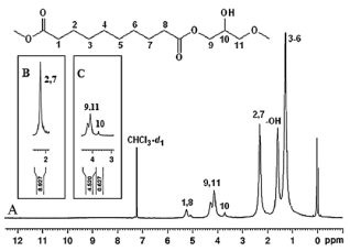
Figure 1. Confirmation of PGS synthesis by comparing H-NMR spectra obtained from PGS synthesis.
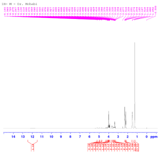
Figure 2. Confirmation of the synthesis of PGS of the present study, based on the results of the H-NMR spectrum obtained from the synthesis of PGS.
3.2. SEM Test
The success rate in producing fibrous scaffolds from PCL and PGS polymers in a two-to-one ratio and the presence of 0.1% gelatin by electrospinning is evident in the SEM images of the scaffolds prepared in this study in Figure 3.
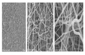
Figure 3. Images from SEM imaging at different magnifications.
It can be seen that the produced fibers are fully intertwined and produce a relatively dense scaffold, which can affect the final properties of the scaffold. It is also clear from high-magnification micrographs that these fibers have a diameter of less than 1 μm.
To more accurately examine the size distribution of the produced microfibers, Image J image processing software was used, and the resulting histogram is shown in Figure 4.
According to Figure 4, it is clear that the fiber diameter size distribution was non-uniform and the largest number of produced microfiber diameter sizes were in the range of 0.2 to 0.4 μm. For a more detailed examination, the statistical parameters extracted from this measurement are reported in Table 1.
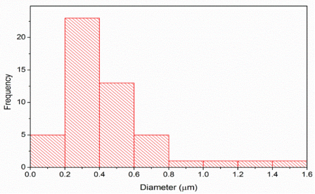
Figure 4. Histogram of the size distribution of the diameter of microfibers related to fibers produced by electrospinning containing PCL and PGS polymers in a ratio of two to one in the presence of 0.1% by weight of gelatin.
Table 1. Statistical parameters extracted from measurements of the diameter size distribution of microfibers related to fibers produced by electrospinning containing PCL and PGS polymers in a ratio of two to one in the presence of 0.1% by weight of gelatin.

Largest diameter (μm)

Smallest diameter (μm)

Average fiber diameter size (μm)

Average fiber diameter size (μm)

Number of measurements

Sample

1.5

0.16

0.28

0.43

50

PCl/PGS/gel

According to Table 1, it is observed that the average diameter of the produced fibers was 0.28 ± 0.48 μm. It is also clear from this measurement that the smallest and largest diameters observed were 0.16 and 1.5 μm, respectively. This non-normal size distribution with a standard deviation of more than half the average value could indicate the relative heterogeneity of the produced fiber.
3.3. FTIR
FTIR tests were performed for PGS, PCL, and gelatin, as well as the final scaffold composition (PGS/PCL/gelatin), and the results are shown in Figures 5-8, respectively. In these graphs, the vertical axis represents the percentage of absorption intensity and the horizontal axis represents the wavelength. In Figure 5, the peaks observed at wave numbers 3466 cm-1, 2855 cm-1, and 2929 cm-1 are respectively related to the stretching vibration of O-H bonds, and the symmetric and asymmetric stretching vibration of C-H bonds (in the form of (-CH2) structures. The intense peak seen at wave number 1736 cm-1 is related to the stretching vibration of C=O double bonds in the PGS polymer. In addition, the symmetric and asymmetric stretching vibration of C-O and C-O-C bonds in the crystalline and non-crystalline structures of PGS has created multiple bonds in the wave number range of 1043 cm-1 to 1162 cm-1. The bending vibration of C-H bonds has also led to various absorption peaks in the wave number range of 1379 cm-1 to 1461 cm-1. Also, the peaks located at 946 cm-1 and 1162 cm-1 723 are also due to the rocking and dancing vibrations of C-H bonds, respectively . In the spectrum obtained from the PCL polymer in Figure 6, the peaks located at wave numbers 2862 cm-1 and 2944 cm-1 are due to the symmetric and asymmetric stretching vibrations of C-H bonds (in the form of (-CH2) structures, respectively. The intense peak related to the stretching vibration of the C=O double bonds, which is due to the ketone group present in PCL, is also located at wave number 1729 cm-1. In addition, the peaks located at wave numbers 1297 cm-1, 1237 cm-1 and 1162 cm-1 are related to the stretching vibration of C-C bonds, the asymmetric stretching vibration of C-O-C bonds and the symmetric stretching vibration of the same bond in the crystal structure of the polymer, respectively. The bending vibration of C-H bonds also produced various absorption peaks in the range of wave numbers from 1364 cm-1 to 1468 cm-1. Also, the peaks located at wave numbers from 1103 cm-1, 1043 cm-1, 961 cm-1 and 730 cm-1 are due to the stretching vibration of C-O-C bonds and the stretching vibration of C-C bonds in the non-crystalline structure of PCL, and the rocking and dancing vibrations of C-H bonds, respectively . According to Figure 7, the peaks observed at wave numbers 3294 cm-1, 3399 cm-1, 2892 cm-1, 2944 cm-1 and 3048 cm-1 are related to the stretching vibrations of O-H and N-H bonds, the symmetric and asymmetric stretching vibrations of C-H bonds and the stretching vibration of bonds connected to aromatic rings, respectively. The stretching vibration of carboxyl and carbonyl bonds (C=O), and the stretching vibration of C-N bonds in primary amine groups present in gelatin also created absorption peaks at wave numbers 1640 cm-1 and 1535 cm-1, respectively. Also, peaks in the range of wave numbers 1334 cm-1 to 1446 cm-1 have emerged due to the bending vibrations of C-H bonds. The observation of absorption peaks at 1238 cm-1 and 11070 cm-1 indicates the presence of C-O and C-O-C bonds in the gelatin used. The peak resulting from the dancing vibrations of C-H bonds in CH2 groups is also located at wave numbers 738 cm-1 and 700 cm-1 . In Figure 8, the symmetric and asymmetric stretching vibration of C-H bonds in CH2 and CH3 groups has created several absorption peaks in the wave number range of 2800 cm-1 to 3000 cm-1. The intense peak related to the stretching vibration of C=O double bonds in the composite under study is located at wave number 1729 cm-1, which is related to this bond in all three PCL, PGS and gelatin compositions. The peaks observed in the range of 2800 cm-1 to 3000 cm-1 wave number are also related to the bending vibration of C-H bonds and the stretching vibration of C-O bonds in the two polymers used as well as gelatin. Also, the intense peak located at about 1215 cm-1 is related to the asymmetric stretching vibration of C-O-C bonds; and the peaks due to the dancing vibrations of C-H bonds also appeared at the wave numbers of 1745 cm-1 and 1670 cm-1. Therefore, the presence of all three compounds of PCL, PGS and gelatin in this composite can be proven from the results of the FTIR test .
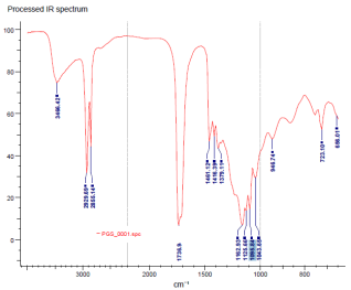
Figure 5. FTIR test result for PGS polymer.
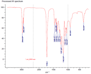
Figure 6. FTIR test result for PCL polymer.
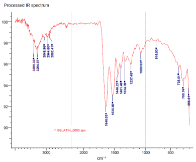
Figure 7. FTIR test result for gelatin polymer.
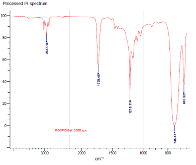
Figure 8. FTIR spectrum for fibrous scaffold (PGS/PCL/gelatin).
3.4. Investigating the Hydrophilicity of Scaffolds Using the Water Contact Angle Test
Figure 9 was prepared to investigate the water contact angle of the final fabricated scaffold (PGS/PCL/gelatin) at times of 10, 20 and 30 seconds. The results show that with the passage of time, the water contact angle has decreased significantly, indicating the hydrophilicity of the fabricated scaffold. These findings are also consistent with the chemical structure of PGS and the presence of hydroxyl functional groups that are responsible for the hydrophilicity of this polymer.
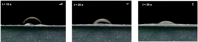
Figure 9. Water contact angle images for electrospun scaffold (PGS/PCL/gelatin) at times of 10, 20 and 30 seconds. The water droplet contact angle has decreased significantly with increasing time. The decrease in the water droplet contact angle means that the produced composite is hydrophilic. In other words, due to the presence of polar groups such as -OH and -NH in the chemical structure of PGS polymer and gelatin, the hydrogen bond formed between these groups and water molecules causes the water droplet to spread on the composite and therefore reduces the contact angle over time.
3.5. Evaluation of Scaffold Mechanical Properties
Uniaxial tensile testing was performed on three samples of the final scaffold. The position of the fibrous scaffold during the tensile test is depicted in this figure. Fortunately, the mechanical properties of the PGS, PCL and gelatin composite are very favorable compared to the PCL/PGS composite and the PGS/gelatin composite previously investigated by other researchers.
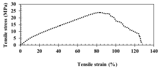
Figure 10. Result of tensile test in the form of stress-strain curve.
Table 2. Quantitative results from tensile testing of manufactured scaffolds.

Toughness (MPa.m0.5)

Failure strain (%)

Failure stress (MPa)

Uniform strain (%)

Tensile strength (MPa)

Young's modulus (MPa)

SAMPLE

1859.43

123.7

8.45

85.6

23.8

0.254

PCL/PGS/Gel

In Table 2, the slope of the part of the curve where the stress changes linearly with strain is equivalent to Young's modulus, and the stress and strain at the maximum point of this curve represent the tensile strength and uniform strain, respectively. Also, the stress and strain at failure are the points after which the stress decreases abruptly and at a constant strain. The toughness is also obtained from the area under the stress-strain curve.
Evaluation of Cell Viability on Scaffolds
To investigate the toxicity of the scaffold (PGS/PCL/gelatin) and to evaluate the viability of cells cultured on it, the MTT test was performed. No significant difference in the rate of cell metabolism, which is an indicator of cell growth and proliferation, was observed in any of the tested times in the experimental and control groups (P-value>0.05); however, on different days and with the lengthening of the cell culture time, the rate of cell metabolism increased significantly in both the control and experimental groups (P-value<0.05) (Figure 10).
4. Conclusion
In previous studies, favorable results were obtained from the use of PGS and PCL polymers in the treatment of heart valve disease; also, a bone scaffold made of PCL and gelatin has been made through electrospinning, which has the necessary strength for use. In this study, an attempt was made to construct an electrospun fibrous scaffold made of PGS, PCL, and gelatin polymers, and to investigate endothelial differentiation by VEGF growth factor on this scaffold in a laboratory environment. Mesenchymal stem cells were cultured on the scaffold and exposed to 50 mg of VEGF to examine the expression of specialized markers CD31 and VEGF-R2.
Environmental conditions such as temperature, air flow, and humidity affect the quality of the fiber obtained from electrospinning, and their continuous control during the process is essential. Another effective factor in the electrospinning process is the parameters related to the solvent. When the polymers dissolve in the solvent, dipoles and ions are created that affect the quality of electrospinning. To solve this problem, the solvent must have a certain concentration to make it easy to control the dipole quality of the material. Also, the parameters of the device, including the type of needle, the distance to the collector, the voltage and the speed, have a direct effect on the characteristics of the final fiber. As the needle leaves the collector head, the surface tension force prevents this action, and whenever the dipole force caused by the electric field exceeds the tension force, the droplet is thrown. Then, the droplet must move forward in a spiral, which is called a Taylor cone; if the spiral movement is controlled in real time with SEM, the conditions are monitored in real time, and if the results are correct, the work continues with the same conditions.
The synthesis of PGS in this study was confirmed after observing the desired functional groups in the NMR test. The formation of polymer particles was also confirmed by measuring the weight of the PGS polymers using the GPC test. Examination of the fiber structure with SEM indicates the success of the electrospinning process. According to the FTIR results and the detection of specific bonds by comparing the characteristic peaks, the presence of all three compounds PGS, PCL and gelatin in the composite was proven. The findings from the investigation of the hydrophilicity of the scaffold or the water contact angle test are consistent with the chemical structure of PGS and the presence of hydroxyl functional groups responsible for the hydrophilicity of this polymer. After the axial tensile test of the scaffold, it was observed that the mechanical properties of the PGS, PCL and gelatin composites were improved compared to the PCL/PGS composite and the PGS/gelatin composite that were investigated by other researchers. The viability of cells cultured on the plate using the MTT test on the second, fourth and sixth day of culture yielded favorable results, indicating the low toxicity of the scaffold used. Also, cell attachment to the scaffold was examined using SEM images and imaging using Dapi staining, both of which indicate proper and efficient cell adhesion. Finally, the expression levels of specialized endothelial markers in each experimental group were measured using the qPCR test and compared with the expression levels of the GAPDH marker in the same group; these results indicate a significant increase in the expression of the two genes CD31 and VEGF-R2.
Abbreviations

PCL

Polycaprolactone

Conflicts of Interest
The authors declare no conflicts of interest.
References
[1] C. B. Weinberg, and E. Bell, “A blood vessel model constructed from collagen and cultured vascular cells”, Science, 231(4736): pp. 397-400, 1986.
[2] L. Niklason, et. al., “Functional arteries grown in vitro”, Science, 284(5413): pp. 489-493, 1999.
[3] K. Shimizu, et. al., “Effective cell-seeding technique using magnetite nanoparticles and magnetic force onto decellularized blood vessels for vascular tissue engineering”, Journal of bioscience and bioengineering, 103(5): pp. 472-478, 2007.
[4] F. W. Sutherland, et. al., “From stem cells to viable autologous semilunar heart valve”, Circulation, 111(21): pp. 2783-2791, 2005.
[5] J. Oswald, et. al., “Mesenchymal stem cells can be differentiated into endothelial cells in vitro”, Stem cells, 22(3): pp. 377-384, 2004.
[6] S. W. Cho, et. al., “Small-diameter blood vessels engineered with bone marrow-derived cells”, Annals of surgery, 241(3): p. 506, 2005.
[7] H. Wang, et. al., “Shear stress induces endothelial differentiation from a murine embryonic mesenchymal progenitor cell line”, Arteriosclerosis, thrombosis, and vascular biology, 25(9): pp. 1817-1823, 2005.
[8] D. Orlic, and J. M. Hill and A. E. Arai, “Stem cells for myocardial regeneration”, Circ. Res., 91(12): pp. 1092 1102, 2002.
[9] M. Majka, and M. Sułkowski, and B. Badyra, and P. Musiałek, “Concise review: mesenchymal stem cells in cardiovascular regeneration: emerging research directions and clinical applications”, Stem cells translational medicine, 6(10): pp. 1859-67, 2017.
[10] G. Zhao, et. al., “Recent Advances in Electrospun Nanofibrous Scaffolds for Cardiac Tissue Engineering”, Adv. Funct. Mater., Vol. 25: 5726-5738, 2015.
[11] V. J. Chen, P. X. Ma, “Polymer Phase Separation: In Scaffolding in tissue engineering”, edited by P. X. Ma and J. Elisseeff, Florida: CRC Press LLC, 2006, pp. 125 - 136.
[12] Z. M. Huang, and Y. Z. Zhang, and M. Kotaki, and S. Ramakrishna, “A review on polyme nanofibers by electrospinning and their applications in nanocomposites”, Compos. Sci. Technol., Vol. 63, pp. 2223-2253, 2003.
[13] W. J. Li, and R. M. Shanti, and R. S. Tuan, “Electrospinning Technology for Nanofibrous Scafffolds in Tissue Engineering”, Nanotechnologies for the Life Sciences, Vol. 9.
[14] Y. Ikada, “Interfacial Biocompatibility: In'Polymers of Biological and Biomedical Significance”, ACS Symposium series 540, edited by S. W. Shalaby and Y. Ikada and R. Langer and J. Williams, Washington DC: American Chemical Society, 1994, pp. 35 - 48.
[15] M. Kharaziha, et. al., “PGS: Gelatin nanofibrous scaffolds with tunable mechanical and structural properties for engineering cardiac tissues”, Biomaterials, 34(27): pp. 6355-6366, 2013.
[16] N. Masoumi, et. al., “Electrospun PGS: PCL microfibers align human valvular interstitial cells and provide tunable scaffold anisotropy”, Advanced healthcare materials, 3(6): pp. 929-939, 2014.
[17] M. E. Hoque, and Y. L. Chuan, and I. Pashby, “Extrusion based rapid prototyping technique: an advanced platform for tissue engineering scaffold fabrication”, Biopolymers, pp. 83-93, 2011.
[18] D. W. Hutmacher, et. al., “Mechanical properties and cell cultural response of polycaprolactone scaffolds designed and fabricated via fused deposition modeling”, J. Biomed. Mater. Res. J., Vol. 55: p. 203, 2001.
[19] D. Hernandez, et. al., “Electrical Stimulation Promotes Cardiac Differentiation of Human Induced Pluripotent Stem Cell”, Stem Cells International, 2015.
[20] K. Tuzlakoglu, and C. M. Alves, and J. F. Mano, and R. L. Reis, “Production and characterization of chitosan fibers and 3-D fiber mesh scaffolds for tissue engineering applications”, Macromol. Biosci., Vol. 4, pp. 811-819, 2004.
[21] M. Rodrigues, and L. G. Griffith, and A. Wells, “Growth factor regulation of proliferation and survival of multipotential stromal cells”, Stem cell research & therapy, 1(4): p. 32, 2010.
[22] K. Holmes, et. al., “Vascular endothelial growth factor receptor-2: structure, function, intracellular signalling and therapeutic inhibition”, Cellular Signalling, 19(10): pp. 2003-2012, 2007.
[23] C. Ikebe, and K. Suzuki, “Mesenchymal stem cells for regenerative therapy: optimization of cell preparation protocols”, BioMed research international, 2014.
[24] F. Berthiaume, M. L. Yarmush, “Fundamentals of Tissue Engineering; Principles and Applications in Engineering Series, edited by B. Palsson and J. A. Hubbell and R. Plonsey and J. D. Bronzino) Florida: CRC Press LLC, 2003, pp. 8-1.
[25] E. S. M. Ng, and N. W. C. Chora, and D. F. Lewis, and O. Hindsgaul, and D. Schriemer, “Frontal affinity chromatography-mass spectrometry”, Nat. Protoc., Vol. 2, pp. 1907-1917, 2007.
[26] Zh. Weilie, “Scanning Microscopy for Nanotechnology Techniques & Applications”, New Orleans: University of New Orleans Press, 2006.
[27] W. E. Teo, and S. Ramakrishna, “A Review on Electrospinning Design and Nanofibre Assem-blies”, Nanotechnology, Vol. 17, 2006.
[28] D. Roylance, “Stress-strain curves”, Massachusetts: Massachusetts Institute of Technology study, Cambridge, 2001.
[29] P. W. Sylvester, “Optimization of the tetrazolium dye (MTT) colorimetric assay for cellular growth and viability”, Methods in molecular biology, 2011.
[30] D. E. Discher, and P. Janmey, and Y. L. Wang, "Tissue cells feel and respond to the stiffness of their substrate", Science, 310 (5751): pp. 1139-43, November, 2005.
[31] S. Bodakhe, et. al, “Injectable photocrosslinkable nanocomposite based on poly (glycerol sebacate) fumarate and hydroxyapatite: Development, biocompatibility and bone regeneration in a rat calvarial bone defect model,” Nanomedicine, Vol. 8, No. 11, pp. 1777-1795, 2013.
[32] H. M. Aydin, and K. Salimi, and Z. M. Rzayev, and E. Pişkin, “Microwave-assisted rapid synthesis of poly (glycerol-sebacate) elastomers. Biomaterials Science, Vol. 1, No. 5: pp. 503-9, 2013.
[33] Q. Liu, et. al., “Structure and properties of thermoplastic poly (glycerol sebacate) elastomers originating from prepolymers with different molecular weights”, Journal of applied polymer science, Vol. 104, No. 2: pp. 1131-7, 2007.
[34] M. R. Jung, et. al., “Validation of ATR FT-IR to identify polymers of plastic marine debris, including those ingested by marine organisms,” Mar. Pollut. Bull., Vol. 127, pp. 704-716, January, 2018.
[35] T. Elzein, et. al., “FTIR study of polycaprolactone chain organization at interfaces,” J. Colloid Interface Sci., Vol. 273, No. 2, pp. 381-387, 2004.
[36] S. Hermanto, and L. O. Sumarlin, and W. Fatimah, “Differentiation of Bovine and Porcine Gelatin Based on Spectroscopic and Electrophoretic Analysis Antihypertensive bioactive peptide from food resources View project,” J. Food Pharm. Sci, Vol. 1, pp. 68-73, 2013.
[37] A. A. Javidparvar, and R. Naderi, and B. Ramezanzadeh, “Non-covalently surface modification of graphene oxide nanosheets and its role in the enhancement of the epoxy-based coatings` physical properties,” Colloids Surfaces A Physicochem. Eng. Asp., Vol. 602, p. 125061, October, 2020.
[38] S. Sant, and C. M. Hwang, and S. H. Lee, and A. Khademhosseini, “Hybrid PGS-PCL microfibrous scaffolds with improved mechanical and biological properties,” J. Tissue Eng. Regen. Med., Vol. 5, No. 4, pp. 283-291, April, 2011.
Cite This Article
  • APA Style

    Kamalijoo, S., Ranjbar, K., Lotfizadeh, D., Nazari, A. (2025). In Vitro Endothelial Differenthelial Assessment on Polyglycerol Sebacate / Polycaprolactone /Gelatin Electospun Scaffold. American Journal of Chemical and Biochemical Engineering, 9(1), 38-47. https://doi.org/10.11648/j.ajcbe.20250901.13

    Copy | Download

    ACS Style

    Kamalijoo, S.; Ranjbar, K.; Lotfizadeh, D.; Nazari, A. In Vitro Endothelial Differenthelial Assessment on Polyglycerol Sebacate / Polycaprolactone /Gelatin Electospun Scaffold. Am. J. Chem. Biochem. Eng. 2025, 9(1), 38-47. doi: 10.11648/j.ajcbe.20250901.13

    Copy | Download

    AMA Style

    Kamalijoo S, Ranjbar K, Lotfizadeh D, Nazari A. In Vitro Endothelial Differenthelial Assessment on Polyglycerol Sebacate / Polycaprolactone /Gelatin Electospun Scaffold. Am J Chem Biochem Eng. 2025;9(1):38-47. doi: 10.11648/j.ajcbe.20250901.13

    Copy | Download

  • @article{10.11648/j.ajcbe.20250901.13,
      author = {Shima Kamalijoo and Kimia Ranjbar and Donya Lotfizadeh and Atousa Nazari},
      title = {In Vitro Endothelial Differenthelial Assessment on Polyglycerol Sebacate / Polycaprolactone /Gelatin Electospun Scaffold
    },
      journal = {American Journal of Chemical and Biochemical Engineering},
      volume = {9},
      number = {1},
      pages = {38-47},
      doi = {10.11648/j.ajcbe.20250901.13},
      url = {https://doi.org/10.11648/j.ajcbe.20250901.13},
      eprint = {https://article.sciencepublishinggroup.com/pdf/10.11648.j.ajcbe.20250901.13},
      abstract = {Due to the prevalence of cardiovascular diseases and their growing trend throughout the world, including Iran, efforts to treat these types of diseases have increased. There are lots of therapies for restoring cardiovascular function include organ transplants, reconstructive surgery, the use of mechanical or artificial devices, and the use of metabolic products. Although these methods are commonly used, they cause problems due to donor limitations, such as biocompatibility, infection, and tissue rejection by the patient. Meanwhile, vascular tissue engineering with the aim of building biocompatible and efficient vessels to replace lost vessels has created high hopes for the treatment of lesions. In this article, we used polyglycerol sebacate polymer (PGS) due to the properties such as high biocompatibility, good cell adhesion, controllable degradation rate and desirable mechanical properties, and combined it with polycaprolactone polymer (PCL) and gelatin to fabricate 3D scaffolds by electrospinning method. We also added vascular endothelial growth factor and then analysed the endothelial differentiation of mesenchymal stem cells. The expression of CD31 and VEGF-R2 genes has been measured by qPCR method, which revealed reasonable results.
    },
     year = {2025}
    }
    

    Copy | Download

  • TY  - JOUR
    T1  - In Vitro Endothelial Differenthelial Assessment on Polyglycerol Sebacate / Polycaprolactone /Gelatin Electospun Scaffold
    
    AU  - Shima Kamalijoo
    AU  - Kimia Ranjbar
    AU  - Donya Lotfizadeh
    AU  - Atousa Nazari
    Y1  - 2025/05/29
    PY  - 2025
    N1  - https://doi.org/10.11648/j.ajcbe.20250901.13
    DO  - 10.11648/j.ajcbe.20250901.13
    T2  - American Journal of Chemical and Biochemical Engineering
    JF  - American Journal of Chemical and Biochemical Engineering
    JO  - American Journal of Chemical and Biochemical Engineering
    SP  - 38
    EP  - 47
    PB  - Science Publishing Group
    SN  - 2639-9989
    UR  - https://doi.org/10.11648/j.ajcbe.20250901.13
    AB  - Due to the prevalence of cardiovascular diseases and their growing trend throughout the world, including Iran, efforts to treat these types of diseases have increased. There are lots of therapies for restoring cardiovascular function include organ transplants, reconstructive surgery, the use of mechanical or artificial devices, and the use of metabolic products. Although these methods are commonly used, they cause problems due to donor limitations, such as biocompatibility, infection, and tissue rejection by the patient. Meanwhile, vascular tissue engineering with the aim of building biocompatible and efficient vessels to replace lost vessels has created high hopes for the treatment of lesions. In this article, we used polyglycerol sebacate polymer (PGS) due to the properties such as high biocompatibility, good cell adhesion, controllable degradation rate and desirable mechanical properties, and combined it with polycaprolactone polymer (PCL) and gelatin to fabricate 3D scaffolds by electrospinning method. We also added vascular endothelial growth factor and then analysed the endothelial differentiation of mesenchymal stem cells. The expression of CD31 and VEGF-R2 genes has been measured by qPCR method, which revealed reasonable results.
    
    VL  - 9
    IS  - 1
    ER  - 

    Copy | Download

Author Information
  • Abstract
  • Keywords
  • Document Sections

    1. 1. Introduction
    2. 2. Method
    3. 3. Discussion
    4. 4. Conclusion
    Show Full Outline
  • Abbreviations
  • Conflicts of Interest
  • References
  • Cite This Article
  • Author Information